Overview
Kidney stones are solid crystalline deposits that can form in the urinary system when the chemical balance of urine changes. Some stones are small and may go unnoticed, while others can obstruct urine flow and create observable effects in the kidneys or ureter.
This article provides an educational explanation of kidney stones, including their formation, types, symptoms, and preventive considerations, using anatomical and physiological insights.
Disclaimer: This article is for educational purposes only. It is not a substitute for professional medical advice. Always consult a healthcare professional for clinical evaluation or guidance.
How the Kidneys Function
The kidneys are paired organs responsible for maintaining the body’s internal chemical balance. Key functions include:
Filtering metabolic waste from the blood
Regulating water and electrolytes (calcium, sodium, potassium)
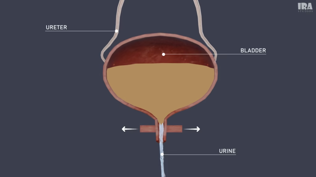
Producing urine as a waste removal medium
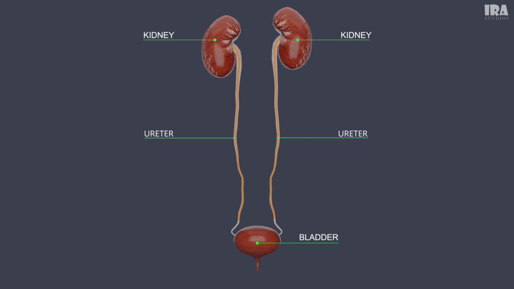
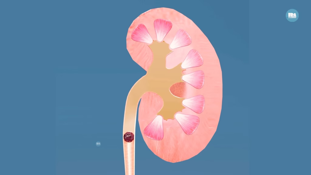
Urine passes through the urinary tract, which includes: (See above image)
Kidneys
Ureters
Urinary bladder
Urethra
Disruptions in urine composition or flow can contribute to crystal formation in the urinary system.
What Are Kidney Stones?
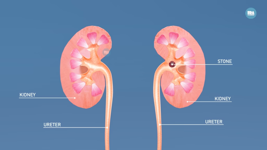
A kidney stone is a hard mass made of crystals that forms anywhere in the urinary tract, including (See above image ):
Kidneys
Ureters
Bladder
Urethra
These stones form when certain substances in urine become too concentrated and stick together, forming crystals. Over time, these crystals grow and harden into stones.
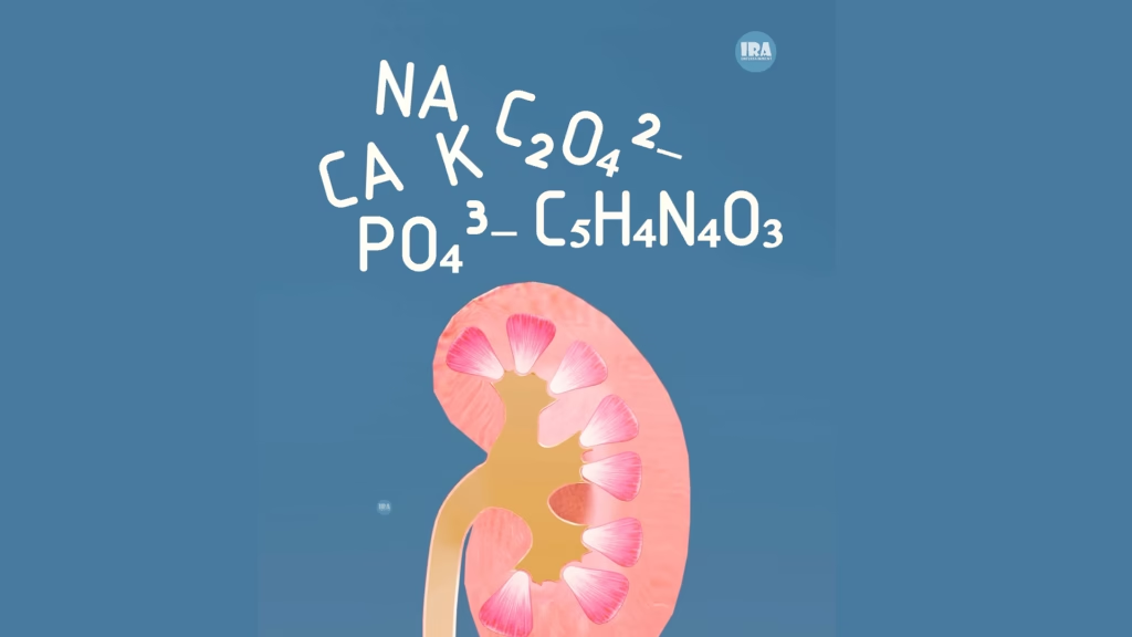
The Chemistry Behind Stone Formation
Urine contains dissolved substances, including:
Calcium
Oxalate
Uric acid
Sodium
Phosphate
Potassium
Normally, these substances are excreted safely. Stones form when concentrations increase or protective factors decrease, allowing crystals to aggregate and grow over time.
How Do Kidney Stones Form?
Kidney stone formation is a gradual, observable process:
Supersaturation
Excess minerals in urine create an environment that favors crystal formation.
Crystal Nucleation
Microscopic crystals begin to form as mineral particles bond, sometimes attaching to kidney tissue.
Crystal Growth and Aggregation
Crystals grow larger and may combine into solid structures.
Stone Maturation
Layering over time results in fully formed kidney stones.
Types of Kidney Stones
Kidney stones are classified by chemical composition:
Calcium Oxalate Stones
Most common (≈80%)
Form when calcium binds with oxalate
Observed in concentrated urine conditions
Uric Acid Stones
Form in acidic urine
Can develop under certain metabolic conditions
Struvite Stones
Associated with urinary system imbalances or chronic infections
Can grow rapidly
Cystine Stones
Rare, genetic stones resulting from cystine accumulation
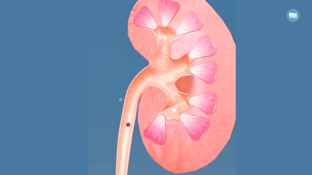
Why Kidney Stones Often Go Undetected
Many people may have a kidney stone without realizing it because these stones can remain completely asymptomatic while they are still inside the kidney. As long as the stone stays in the kidney and does not block the flow of urine, it often causes no pain, discomfort, or noticeable changes in bodily functions. Routine check-ups or imaging studies are often the only way to detect these silent stones.
The real issue begins when the stone moves out of the kidney and enters the ureter, the narrow tube that connects the kidney to the bladder. Even a small stone can cause intense pain, known as renal colic, by obstructing the flow of urine. This can lead to symptoms such as sharp flank pain, blood in the urine, nausea, and frequent urination.
Because kidney stones can remain hidden for months or even years, many people are caught by surprise when the first episode of pain occurs. Understanding this silent nature of kidney stones highlights the importance of preventive measures, such as staying hydrated, maintaining a healthy diet, and monitoring risk factors like family history or metabolic conditions.
Observing Discomfort Mechanisms
The ureter is a narrow, muscular tube that propels urine using rhythmic contractions. When a stone enters:
The ureter stretches mechanically
Its lining may experience irritation from the stone
Muscle contractions intensify to move the obstruction
This process may result in renal colic, which is characterized by wave-like sensations that can extend from the back toward the groin.
Common Symptoms of Kidney Stones
Symptoms vary depending on stone size and location, and may include:
Flank or lower back sensations
Radiating sensations to the abdomen or groin
Observable changes in urine (e.g., color)
Increased urinary frequency
Nausea or vomiting
Fever-like responses in cases of system imbalance
Note: These observations are presented from an anatomical perspective and are not personal medical advice.
Observing Potential Complications
If urine flow is obstructed, the following changes can be seen anatomically:
Swelling of kidney structures (hydronephrosis)
Stress on kidney tissue
Rarely, significant impairment of kidney function
Stone Movement and Management
Yes.
Most stones that are smaller than 5 millimeters in diameter can pass through urine without special medical treatment.
Educational observation:
Drinking plenty of water
Pain management
Monitoring symptoms
The stone gradually moves out of the body naturally.
Observing Larger Stones
Medical intervention may required if:
The stone is too large to pass
Pain is uncontrollable
There is infection
Urine flow is blocked
Treatment options may include:
Medications
Shock wave therapy (lithotripsy)
Endoscopic stone removal
Surgery (rare cases)
Larger stones or persistent observations are generally tracked using clinical imaging techniques. This section emphasizes anatomical and mechanical perspectives rather than personal treatment instructions.
Preventive Considerations
Prevention is always better than treatment.
Simple preventive steps:
Drink enough of water daily
Reduce excess salt intake
Balance calcium consumption (don’t avoid it completely)
Limit foods high in oxalate if prone to stones
Maintain a healthy lifestyle
Frequently Asked Questions
Can kidney stones dissolve naturally?
Certain uric acid stones may dissolve under specific chemical conditions, but most stones require passage through the urinary system or observation in clinical simulations.
Do kidney stones always cause discomfort?
Not always. Stones may remain asymptomatic until urine flow is physically affected.
Can kidney stones recur?
Yes. Awareness of crystal formation factors and preventive considerations can help reduce recurrence.
Key Takeaways
Kidney stones are crystalline structures formed from urinary minerals. Understanding their anatomy, formation, types, and 3D movement can enhance educational awareness and help visualize their impact on the urinary system.
This article is an informational educational resource and does not replace professional medical consultation.
About the Author: This article was prepared by the Ira Studios editorial team. We specialize in creating high-fidelity 3D educational visualizations. Our mission is to bridge the gap between complex anatomy and visual education through deep-dive 3D cross-sections and animations.
Watch the Full 3D Animation
To explore all of these processes in 3D, check out our detailed 3D Animation video.
Watch it in Malayalam!
Want to learn more through visual storytelling? Check out our detailed 3D explanation blog on Understanding Heart Attacks: A 3D Explanation of Causes, Symptoms, and Prevention